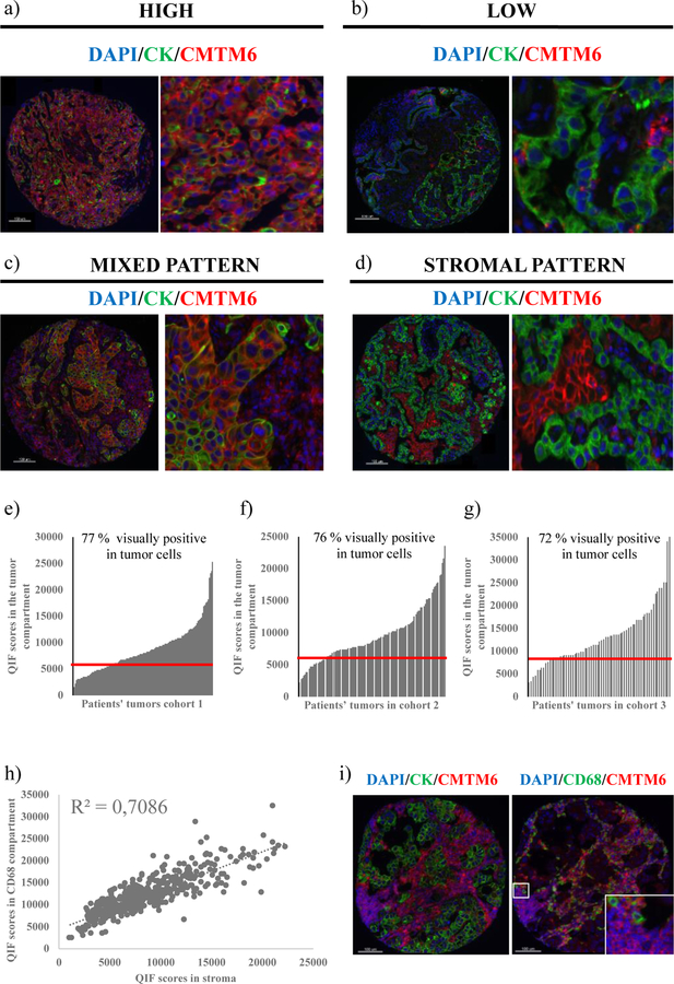
Figure 1. Patterns of CMTM6 expression in human NSCLC tissue.
(a)-(b) Representative cases with high and low CMTM6 expression; (c)-(d) Representative cases with mixed stromal and tumor CMTM6 expression (c) and stroma-predominant expression (d); (e)-(g) Dynamic range of CMTM6 expression in the tumor compartment in the three tested cohorts. Red line represents the visual CMTM6 detection threshold in tumor cells; (h) Correlation between CMTM6 QIF scores in the stromal compartment vs. the CD68 compartment (three cohorts combined, n = 438); (i) Representative image of CMTM6 expression in CD68 positive and CD68 negative cells in the stroma

