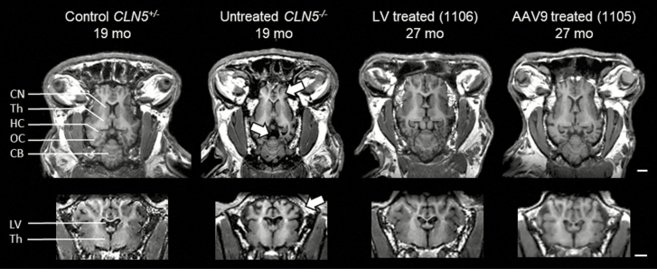
Figure 4.
T1-weighted MRI images showing the preservation of neuroanatomical structures after gene therapy in CLN5−/− sheep. T1-weighted MRI images in the horizontal (top) or coronal (bottom) views from 19-mo old control or affected sheep compared with much older (27 mo old) sheep treated by gene therapy with lentivirus (LV) or single-stranded adeno-associated viral serotype 9 (AAV9) before the development of clinical signs. The treatments protected from the high neuronal atrophy observed in affected animals because either vector preserved neuroanatomical structures, protecting against the profound cortical atrophy (top arrow), prominent ventricular enlargement (center arrow), and cranial thickening (bottom arrow) seen in the untreated CLN5−/− animals. CB = cerebellum; CN = caudate nucleus; HC = hippocampus; LV = lateral ventricles; OC = occipital cortex; Th = thalamus. By permission from Elsevier (original publication: Mitchell et al. (2018), Molecular Therapy, 26(10): 2366–2378).

