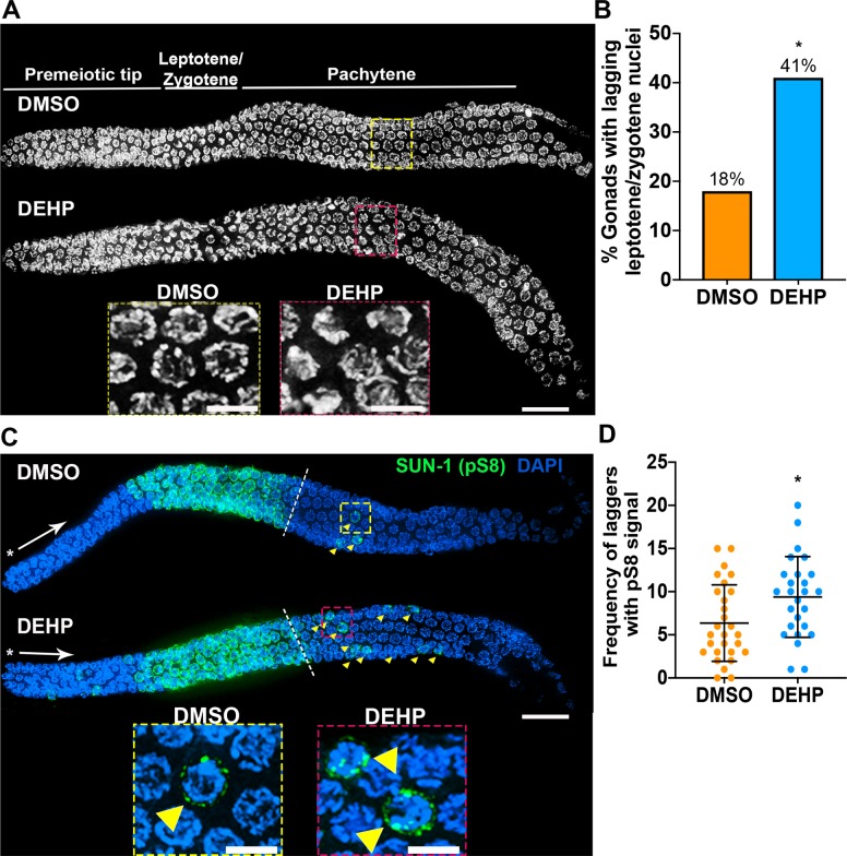
Fig 2. Meiotic progression is altered in the germline of DEHP-exposed hermaphrodites.
(A) High-resolution images of whole mounted gonads from DMSO- or DEHP-exposed worms oriented from left to right. Insets show the presence of transition zone-like nuclei in mid-pachytene for DEHP-exposed gonads suggesting the presence of lagging leptotene/zygotene nuclei. (B) Quantification of gonads with lagging leptotene/zygotene nuclei. Data is from four independent biological repeats for DMSO (n = 39) and DEHP (n = 39; n = number of gonads scored, *P<0.05 by Fisher’s exact test). (C) Whole mounted gonads of chemically-exposed worms immunostained against SUN-1 (pS8) (green) show defects in meiotic progression as manifested by the persistence of pS8+ nuclei (“laggers”, indicated by yellow arrows) into mid to late pachytene. Asterisks indicate the premeiotic tip and white arrows show the direction in which nuclei move proximally in the germline. Hatched white lines demarcate the end of rows with all nuclei showing SUN-1 (pS8) signal (beginning of mid-pachytene). Insets show the presence of pS8+ lagging nuclei in the mid-pachytene regions of DMSO- and DEHP-exposed germlines. (D) Quantification of the number of pS8+ laggers observed in mid and late pachytene per gonad. Bars represent the mean number of laggers ± SD. *P<0.05 by the two-tailed Mann-Whitney test, 95% C.I.; Effect size = 5. Data was obtained from four biological repeats for DMSO (n = 28) and DEHP (n = 28); n = number of gonads scored. Scale bars, 5 μm.

