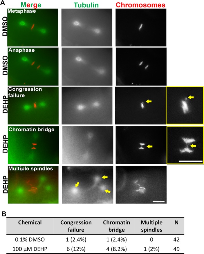
Fig 4. DEHP exposure causes defects in the first embryonic division.
Representative images from time-lapse analysis of the first embryonic division in DMSO- and DEHP-exposed H2B::mCherry; γ-tubulin::GFP; col-121(nx3) worms. (A) DEHP exposure led to chromosomes failing to properly align on the metaphase plate (indicated by yellow arrow in inset), chromatin bridges at the metaphase to anaphase transition (indicated by yellow arrow in inset) and the formation of multiple spindles (indicated by yellow arrows). Chromosomes are shown in red and tubulin in green. Data for 0.1% DMSO (n = 42) and 100 μM DEHP (n = 49) was obtained from five biological replicates; n = total number of embryos scored from 42 and 49 independent animals, respectively. Scale bars, 5 μm. (B) Quantification of defects scored by time-lapse analysis of the first embryonic. A total of 7 out of 49 (14%) embryos displayed defects in the case of DEHP exposures, while only 2 out of 42 (4.8%) embryos showed defects in the case of DMSO. Four of the DEHP-exposed embryos exhibited more than one class of defect (see S1 Table).

