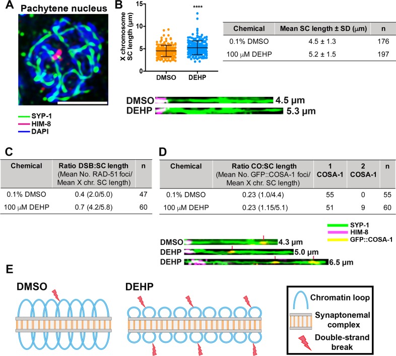
Fig 8. DEHP exposure alters X chromosome SC length and CO designation.
(A) High magnification image of a full-projection of a mid-pachytene nucleus immunostained against the central region component of the SC, SYP-1 (green), and the pairing center end protein HIM-8 (magenta). Scale bar, 2 μm. (B) Scatter plot showing the distribution of X chromosome SC lengths in DMSO- and DEHP-exposed worms. The X chromosome SC length is significantly extended in DEHP-exposed pachytene nuclei compared to DMSO (****P < 0.0001 by the two-tailed Mann-Whitney test, 95% C.I.). Error bars represent mean ± SD. Shown on the right are the mean lengths of the X chromosome SC and the total numbers of nuclei scored (DEHP: n = 197 nuclei from 61 gonads, DMSO: n = 176 nuclei from 53 gonads; data collected from six independent biological repeats). Shown below the graph are X chromosomes, identified based on HIM-8 staining (magenta), traced in 3D along the SYP-1 signal (green) and straightened computationally using Priism 4.7. (C) Mean number of RAD-51 foci scored/mean length (μm) for computationally straightened X chromosomes from the same analysis. This analysis shows an increase in the ratio of DSB:SC length in pachytene nuclei from DEHP-treated worms. n = number of nuclei scored. (D) Mean number of GFP::COSA-1 foci scored/mean length (μm) for computationally straightened X chromosomes from the same analysis. Ratios of CO:SC length were the same for DEHP and DMSO treated worms. However, 2 COSA-1 foci per X chromosome were only detected in DEHP-treated worms and COSA-1 foci levels observed on the X chromosome were significantly higher in DEHP-treated worms compared to DMSO (*P<0.05 by a two-tailed Mann-Whitney test, 95% C.I.). Shown below are computationally straightened X chromosomes with HIM-8 (magenta), SYP-1 (green) and GFP::COSA-1 (yellow). Red vertical lines indicate COSA-1 foci. (E) Schematic illustrating the potential organization of chromatin loops in vehicle alone (DMSO) treated meiotic nuclei (left) compared to DEHP-treated nuclei (right). In the latter, the SC length is increased, which may be associated with decreased loop length and increased numbers of chromatin loops at which meiotic DSBs are proposed to take place.

