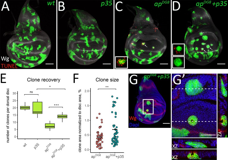
Fig 5. In the absence of apoptosis misspecified clones become bigger and undergo extrusion.
(A-D) Third instar wing discs with wild-type (A), p35-expressing (B), apDG8 (C) and apDG8 p35-expressing clones. The insets in C and D show enlarged images of single representative clones defined by arrows. (E) Clone recovery in the dorsal disc. 9 discs for each genotype were analyzed. (F) Plot shows areas of apDG8 and apDG8 p35-expressing clones. (G-G’) ap mutant clones are eliminated from the dorsal pouch via extrusion when apoptosis is blocked. (G) Third instar wing disc containing apDG8 p35-expressing clones. (G’) Zoom-in of the region defined by the white square in G. The XZ and YZ cross-sections throughout the clones located in the hinge and in the pouch are shown (XZ orientation: the apical side–up; YZ orientation: the apical side—left). Scale bars represent 50μm.

