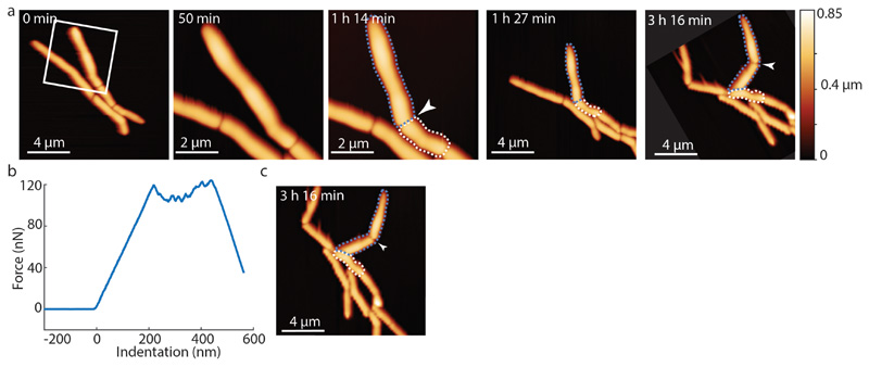
Extended Data Figure S13. Cells forced to undergo premature cleavage continue to grow and divide.
(a) Time lapse AFM height images of cells that continue to grow and divide after cleavage was prematurely induced by application of mechanical stress on the PCF using the AFM cantilever. At 50 minutes, a zoom-in shows the area outlined at 0 minutes (white box). At 1 hour 14 minutes, premature cell cleavage was induced by AFM-mediated application of force on the PCF (white arrow). Between 1 hour 27 minutes and 3 hours 16 minutes, the prematurely cleaved new cell poles elongated and the corresponding sibling cells grew and divided (arrowhead at 3 hours 16 minutes, cropped and rotated to align with previous time points). (b) Force curve corresponding to forced cell cleavage at 1 hour 14 minutes. (c) Non-cropped and non-rotated image of time-lapse image shown in (a) at 3 hours 16 minutes.

