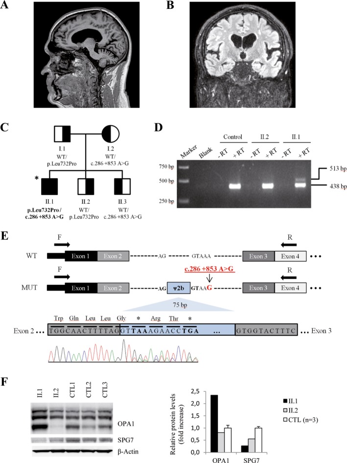
Figure 1.

MRI brain images and functional evaluation in fibroblasts from Patient II.1. (A, B) Brain MRI of subject II.1 showing typical cerebellar atrophy of SPG7 disease (sagittal T2w) (A), and bilateral frontotemporal and right parietal atrophy with grade 1 subcortical leukoencephalopathy (coronal FLAIR) (B). (C) Family pedigree and genotype data for the SPG7 variants. Symbols: square, male; circle, female; filled, affected individual; half‐filled, clinically healthy carriers; asterisk, individual sequenced by WES/WGS. Variants found in SPG7 are shown below each symbol. WT: wild‐type. (D) Agarose gel electrophoresis of RT‐PCR products showing an additional fragment (513 bp) in patient II.1. No products were observed in negative RT controls or water controls (blank). A healthy family member (II.2) not carrying the c.286 + 853A>G variant and an unrelated individual were used as healthy controls. (E) Consequence of the c. 286 + 853 A> G variant. WT: structure of wild‐type SPG7 transcript (exons 1–4). MUT: structure of SPG7 transcript generated by the c. 286 + 853 A> G variant. The pseudoexon results from a cryptic donor splice site activation upstream from the c.286 + 853A>G mutation. The in‐frame inclusion of 75 nucleotides of intron 2 (blue, pseudoexon ᴪ2b) translates into premature stop codons at positions 1 and 4 downstream. Arrows indicate primers used for RT‐PCR. cDNA Sanger sequencing shows two populations of mRNA, one corresponding to pseudoexon inclusion and the other to exon 3. (F) Western blot of OPA1 and SPG7 proteins for human fibroblasts of individuals II.1 (patient), II.2 (healthy brother) and controls (n = 3). Right, quantification of western blot bands. Data represented as mean ± SD
