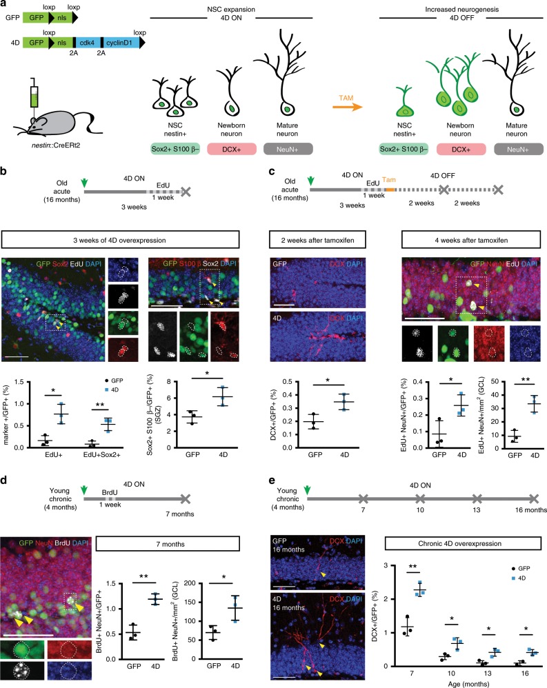
Fig. 1. 4D increases NSC expansion and neurogenesis throughout life.
a Scheme depicting the GFP and 4D viral constructs and experimental approach to increase NSC expansion and neurogenesis. Temporal control of transgenes expression was achieved by delivering lentiviruses to nestin::CreERt2 mice (4D ON) with tamoxifen being later used to delete the LoxP-flanked 4D cassette (4D OFF) together with the nuclear localisation signal (nls) of GFP (or only the nls in control, GFP viruses). Effects on cell types, nuclear to cytoplasmic redistribution of GFP and neurogenic markers used in this study are depicted in the cartoon simplifying the neurogenic lineage from stem and progenitor cells (NSC) to newborn and mature neurons. b–e Experimental layouts (top), fluorescence pictures and quantifications (middle/bottom) of cell types identified upon immunohistochemistry for neurogenic markers (as indicated) in the DG or subgranular zone (SGZ) of mice injected with GFP or 4D viruses and analysed at different times later, as indicated. Insets (dashed boxes, b–d) are magnified and examples of cells scored indicated (arrowheads or dotted lines). Scale bars = 50 μm. Data represent mean ± SD as the proportion of cells scored within the GFP+ infected population or total numbers of cells per area (mm2) in control (black) or 4D (blue) mice; N = 3; n > 2000 (b–e) and >1000 (b, right); error bars = SD (except for e, GFP, 16 months: N = 2; bar = SEM). *p < 0.05; **p < 0.01 assessed by unpaired Student’s t test.

