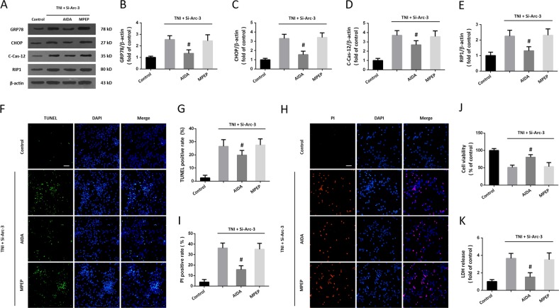
Fig. 7. Role of mGluR1 pathway in Arc knockdown-induced ER stress and necroptosis.
a–e Western blot a and quantification b–e show that the increased expression of GRP78 b, CHOP c, cleaved-caspase-12 d, and RIP1 e induced by Arc knockdown after TNI were partially prevented by AIDA, but not by MPEP. f, g TUNEL staining f and quantification g show that the enhanced neuronal apoptosis induced by Arc knockdown after TNI was attenuated by AIDA, but not by MPEP. h, i PI staining h and quantification i show that the increased number of PI-positive cells induced by Arc knockdown after TNI was inhibited by AIDA, but not by MPEP. j Cell viability assay shows that the decreased cell viability induced by Arc knockdown after TNI was alleviated by AIDA, but not by MPEP. k LDH release assay shows that the increased LDH release induced by Arc knockdown after TNI was partially prevented by AIDA, but not by MPEP. Error bars indicate SEM (n = 6). *p < 0.05 vs. Control group. #p < 0.05 vs. TNI + Si-Arc-3 group.

