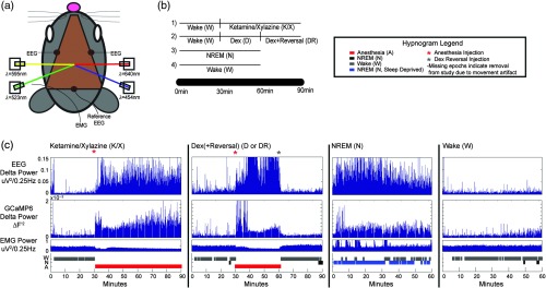
Fig. 1.
Concurrent GCaMP6 fluorescence, OIS imaging, and EEG acquisition system with experimental design. (a) Mouse head schematic of chronically implanted Plexiglass window used for consistent, repeated imaging experiments (brown). Two EEG screws were implanted bilaterally in the skull, with a reference EEG electrode near the cerebellum and EMG wire threaded through the neck as shown. OIS imaging was performed using sequential LED illumination supplied by three LEDs (523, 595, and 640 nm). GCaMP6 fluorescence imaging was performed using a 454 nm LED for excitation. (b) Outline of experimental design. For experiments (1) and (2), 30 min of spontaneous wake data was acquired, injection of the anesthetic followed, with the reversal agent given in run (2) at 60 min to reverse the effects of dex. In runs (3) and (4), 60 min of spontaneous NREM (if sleep deprived) or wake (if not sleep deprived) data was acquired to be scored. (c) Example delta (0.7–3.0 Hz) power time course for EEG and GCaMP6 data with corresponding EMG and hypnogram for one mouse during ketamine/xylazine (K/X), dex (D), dex+reversal (DR), NREM (N), and wake (W).

