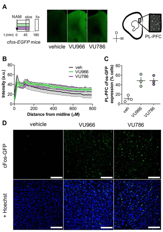
Figure 1. Systemic administration of either mGlu2 or mGlu3 NAM rapidly activates the mouse PFC.
(A) c-Fos-EGFP mice received vehicle, the mGlu2 NAM VU6001966 (VU966, 10 mg/kg), or the mGlu3 NAM VU0650786 (VU786, 30 mg/kg), 45 minutes prior to sacrifice for slice preparation, fixation, and processing for immunohistochemistry. Representative coronal widefield images displaying c-Fos-EGFP expression throughout several medial PFC areas following NAM administration. (C) c-Fos-EGFP expression throughout all layers of the medial PFC. (C) Prelimbic PFC slices from NAM-treated mice displayed robust increases in the proportion of c- Fos-GFP-expressing cells. N = 3 mice per group with 2-4 replicates per mouse. (D) Representative images of c-Fos-GFP expression (top) and co-localization with Hoescht nuclear stain used for the quantification in panel D. Scale bars represent 100 μm.

