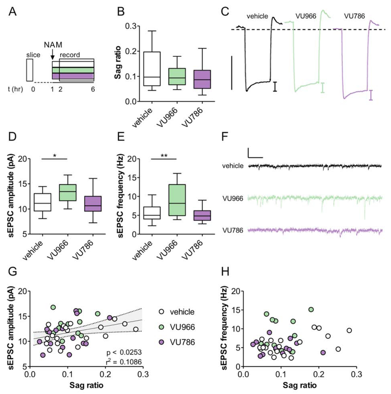
Figure 3. mGlu2 inhibition in acute PFC slices increases excitatory synaptic strength.
(A) PFC slices were prepared and incubated with vehicle, VU6001966 (VU966; 3 μM), or VU0650786 (VU786; 10 μM) for 1-6 hours prior to whole-cell recordings. (B) No effect of slice NAM treatment on hyperpolarization sag (ANOVA main effect of NAM: F2,44 = 1.7, p<0.27, n/N = 12-20/4 cells/mice per group). (C) Representative traces displaying membrane hyperpolarization in response to a 150 pA hyperpolarizing current. Scale bar represents 10 mV. Dashed line indicates −70 mV. (D-E) Incubation with the mGlu2 NAM, but not the mGlu3 NAM, increased sEPSC amplitude (F2,44 = 2.3, p<0.04, n/N = 13-19/4; *: p < 0.05 Bonferonni post-test vs. vehicle) and frequency (F2,43 = 11.98, p<0.0034, n/N = 13-19/4; **: p < 0.01 Bonferonni post-test vs. vehicle). (F) Representative sEPSC traces. Scale bars indicate 20 pA and 100 ms. (G) Positive correlation between sEPSC amplitude and hyperpolarization sag. (H) No correlation between sEPSC frequency and sag ratio.

