Fig. 5.
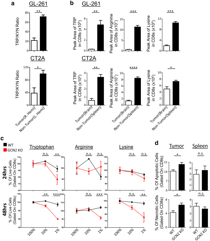
Amino acid deficiency causes necrosis in the absence of GCN2 in CD8+ T-cells in murine glioma tumor models. In a, WT mice were injected with GL-261 (top panel) and CT2A (bottom panel) tumor cells, three mice per group. After 14 days of tumor growth, tumor (R. Hemi) and non-tumor (L. Hemi) hemisphere were isolated and measured for TRP/KYN ratios via LC–MS. In b, WT mice were injected with GL-261 (top panel) and CT2A (bottom panel) tumor cells, six mice per group. After 14 days, mice were euthanized and CD8β+ T-cells were isolated from the tumor (brain) and periphery (spleen), and their metabolites were measured using LC–MS. In c, total T-cells were cultured in serial dilutions of three amino acids, and at 24 and 48 h, cells were lifted and stained with annexin-V and fixable viability dye for flow cytometric analysis. Refer to Supplementary Fig. 7d for the gating strategy. In d, WT and GCN2 KO OT-1 CD8+ T-cells were transferred i.v. into previously inoculated with GL-261 OVA, Rag1 KO mice. Seven days post transfer, mice were euthanized, and tumor-infiltrating lymphocytes were stained with annexin-V and fixable viability dye for flow cytometric analysis. Refer to Supplementary Fig. 7e for the gating strategy. In a, the ratio of normalized peak area of TRP to KYN and in b is the normalized number of the peak area of tryptophan, arginine and lysine in the tumor versus non-tumor. In c, Two-way ANOVA followed by Sidak’s multiple comparisons test was used to calculate significance, n = 3 per group. In d, flow cytometry statistics calculated and shown as percentage positive population, n = 3 per group, representative of two experiments. Unpaired T test analysis was used to calculate significance. p < 0.05*; p < 0.01**; p < 0.001***; p < 0.0001****

