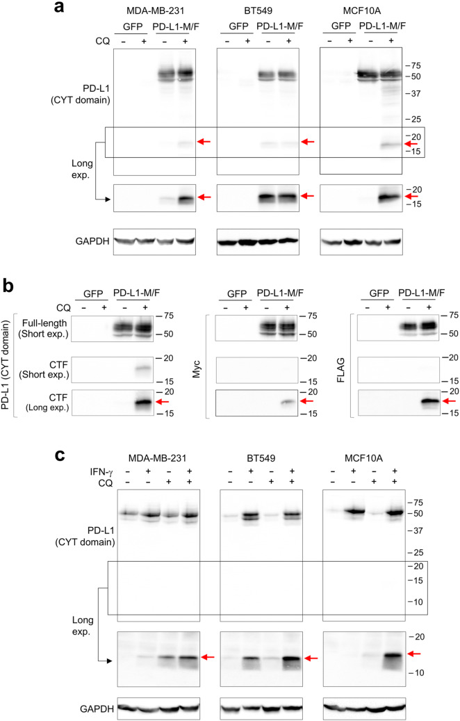
Fig. 4.
Detection of the C-terminal proteolytic fragment of PD-L1. a MDA-MB-231, BT549, or MCF10A cells overexpressing PD-L1-M/F or GFP (control) were incubated for 24 h in the absence or presence of 10 μM chloroquine (CQ). Cell lysates were analyzed by Western blotting using an antibody specific for the cytoplasmic (CYT) domain of PD-L1 (E1L3N). The low molecular weight region of the blot (indicated by the box) is also shown after a longer exposure time. b MDA-MB-231 cell lysates from a were analyzed by anti-PD-L1 (E1L3N), anti-Myc, or anti-FLAG antibody. Short and long exposures of the low molecular weight region are shown. CTF, C-terminal fragment. c Parental MDA-MB-231, BT549, or MCF10A cells were incubated for 48 h in the presence or absence of 10 ng/ml IFN-γ, and then for additional 24 h with 10 μM CQ in the presence or absence of IFN-γ, as indicated. Cell lysates were analyzed by Western blotting using the E1L3N anti-PD-L1 antibody. The low molecular weight region of the blot (indicated by the box) is also shown after a longer exposure time. GAPDH is a gel loading control. Results are representative of three (a) or two (b, c) independent experiments

