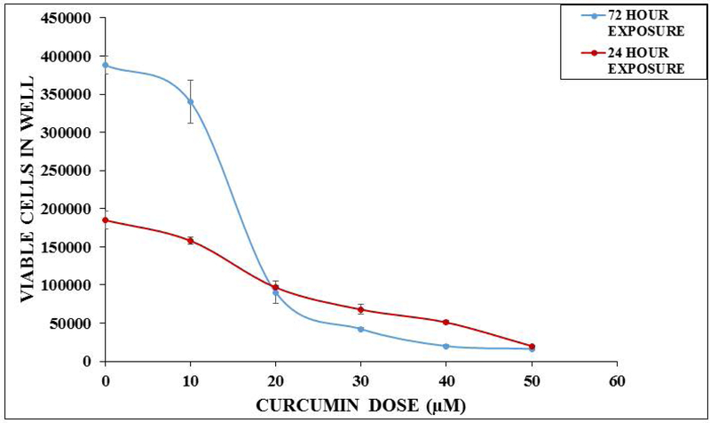
Figure 4.
In vitro static toxicity of curcumin on MDA MB231 (breast) cells. MDA MB231 cells were resuspended at 5 × 10⁻⁴ cells/mL, 1 mL of the suspension was plated into 6 well-plates. After 24 hours, the plates were divided into the following groups: (a) Control (b) 1 μM curcumin (c) 10 μM curcumin (d) 20 μM curcumin (e) 30 μM curcumin (f) 40 μM curcumin (g) 50 μM curcumin. Cell densities were determined using a hemocytometer after 24 hours or 72 hours of curcumin exposure. Data are expressed as standard error of the mean (n = 3 separate experiments). The in vitro LC50 values were calculated to be 22 μM and 15 μM, respectively.

