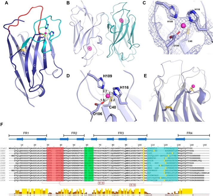
Figure 2.
Structural features of ICab3. A, overall structure of ICab3. CDR1, CDR2, and CDR3 regions are represented by red, green, and cyan, respectively; transparent spheres depict the disulfide bond between Cys26 and Cys103. B, anomalous map (magenta mesh) of ICab3 contoured at 6.0σ showing a strong density in the CDR3 of each chain. C, 2Fo − Fc map contoured at 3.0σ showing Zn2+-coordinating residues in the Zn2+-binding pocket. D, Zn2+ is bound with a tetrahedral coordination geometry, outlined with gray lines; dashes show distances between Zn2+ and coordinating atoms (Å). E, structural superposition of ICab3 (light blue) with another camelid VHH (gray; PDB code 3JBE) showing overlap of the Zn2+-binding region of ICab3 (light blue) with that of the disulfide bond of 3JBE. F, multiple-sequence alignment of ICab3 with other VHH sequences whose structures are known. β-Strands are represented by overhead arrows, whereas CDR1 to -3 are highlighted in the same color as in A. Cysteines that form disulfides have been shown with connectors (-S-S-), whereas the region in CDR3 that harbors additional cysteine is demarcated with a dashed line. Conservation scores are given in the histogram below. 0, least conservation; *, total conservation. −, unassigned region. FR1–4 represent framework regions 1–4 in the VHH.

