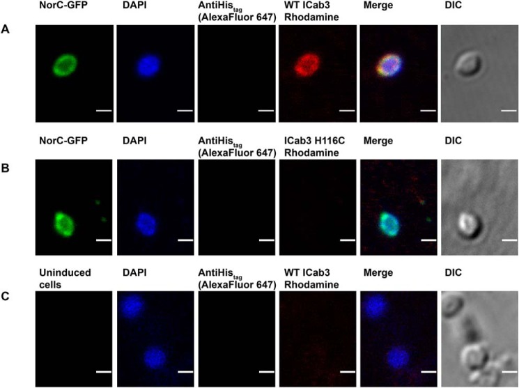
Figure 6.
Confocal imaging showing WT ICab3 binding to the extracellular face of NorC in E. coli spheroplasts. Images were acquired after incubating E. coli spheroplasts expressing NorC-GFP with rhodamine-labeled WT ICab3 (A) and rhodamine-labeled ICab3 H116C (B). C, images acquired with uninduced E. coli spheroplasts incubated with rhodamine-labeled WT ICab3. Total number of cells counted for A = 40, and that for B and C was 15 each in four trials. Scale bar, 1 μm. DIC, differential interference contrast.

