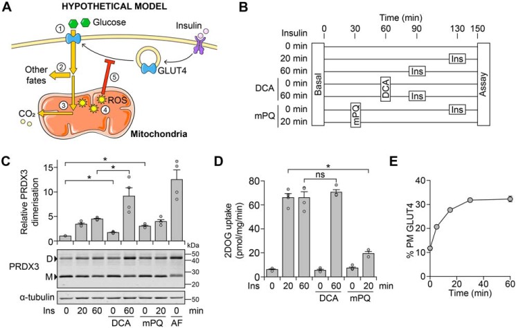
Figure 3.
Glucose-dependent mitochondrial oxidants do not cause insulin resistance. A, schematic depicting how insulin-dependent glucose oxidation could lead to oxidant-induced insulin resistance. Glucose is taken up (1), and a majority is diverted to fates such as lipid or lactate production (2) (21).7 A small portion of glucose is oxidized (3), and although this does not contribute substantially to respiration, it increases mitochondrial ROS levels (4), eventually leading to insulin resistance (5). B–D, 3T3-L1 adipocytes were treated in Medium B with mPQ (20 μm), DCA (1 mm), and insulin (Ins, 100 nm) at the times indicated in B. Cells were also treated with auranofin (AF; 5 μm) at t = 30 min (concurrently with mPQ treatment) in C. Cells in C and D were treated in parallel. Following treatment, cells were either harvested for protein and lysates subjected to Western blotting with antibodies against the indicated proteins (C) or assayed for 2-deoxyglucose (2DOG) uptake under cold conditions (D). In C, densitometric analysis was performed as described in the legend to Fig. 2D. Data are presented as mean ± S.E. (error bars) from four separate experiments. *, p < 0.05; ns, p > 0.05 by two-sample t test; D, dimer; M, monomer. E, 3T3-L1 adipocytes constitutively expressing HA-tagged GLUT4 were incubated in Medium B and stimulated with Ins (100 nm) for the indicated times, after which GLUT4 translocation to the PM was measured. Data are presented as mean ± S.E. from three separate experiments.

