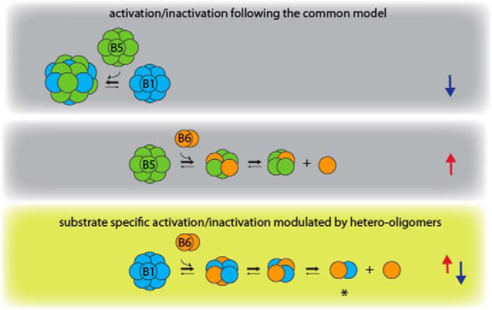
Figure 7.

Model of activity modulation by hetero-oligomerization between HspB1, HspB5, and HspB6. The three different types of sHsp hetero-oligomers show differences in oligomeric size and chaperone activity. Arrows on the right indicate an increase (red) or decrease (blue) in chaperone activity. HspB6 activates HspB5 upon hetero-oligomer formation, whereas the large HspB1–HspB5 hetero-oligomers are inactivated. Thus, these two types of hetero-oligomers follow the common model of chaperon activity regulation (indicated by gray background) by shifting the oligomer distribution to larger oligomers (inactive species) or to smaller oligomers (active species). In contrast, HspB1–HspB6 hetero-oligomers show regulation in both directions in a substrate-dependent manner (olive green background), which seems to be a result of the specific formation of heterodimers. Circuits do not necessarily represent monomers.
