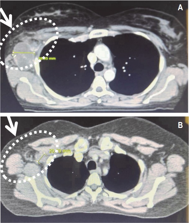
Fig. 2.

Computerized tomography (CT) imaging of the axilla. (A) CT scan of the axilla showing confluent mass in axilla prior to central nervous system (CNS) radiation therapy. (B) CT scan of the axilla showing dissipating tumor following CNS radiation therapy.
