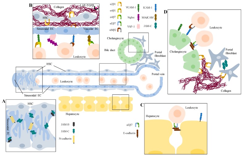
Figure 1.
Overview of hepatic cell interactions mediated by CAMs during liver inflammation and fibrosis. Displayed is a sinusoidal channel near a portal vein and a bile duct. (A) The microvessel is covered by activated hepatic stellate cells (HSCs), which act as mural cells. They interact with each other via N-cadherin and JAM-C homophilic binding and with sinusoidal endothelial cells (ECs) via JAM-B/JAM-C interaction. (B) HSCs are the main producers of fibrotic extracellular matrix (ECM), like collagen I. Tethered to the ECM is LAP-TGFβ. Chemokine-attracted leukocytes get recruited to capillarized sinusoidal ECs by binding via integrins to members of the immunoglobulin (Ig) superfamily of cell adhesion molecules (IgCAMs) or to non-classical CAMs like VAP-1, or they bind to MAdCAM-1 on vascular ECs before they transmigrate the endothelial wall and interact later on with (C) hepatocytes. (D) In biliary disease, portal fibroblasts also get activated, secrete fibrotic ECM and bind to each other via JAM-C. Further, leukocytes attach to cholangiocytes, e.g., via integrin/IgCAM binding.

