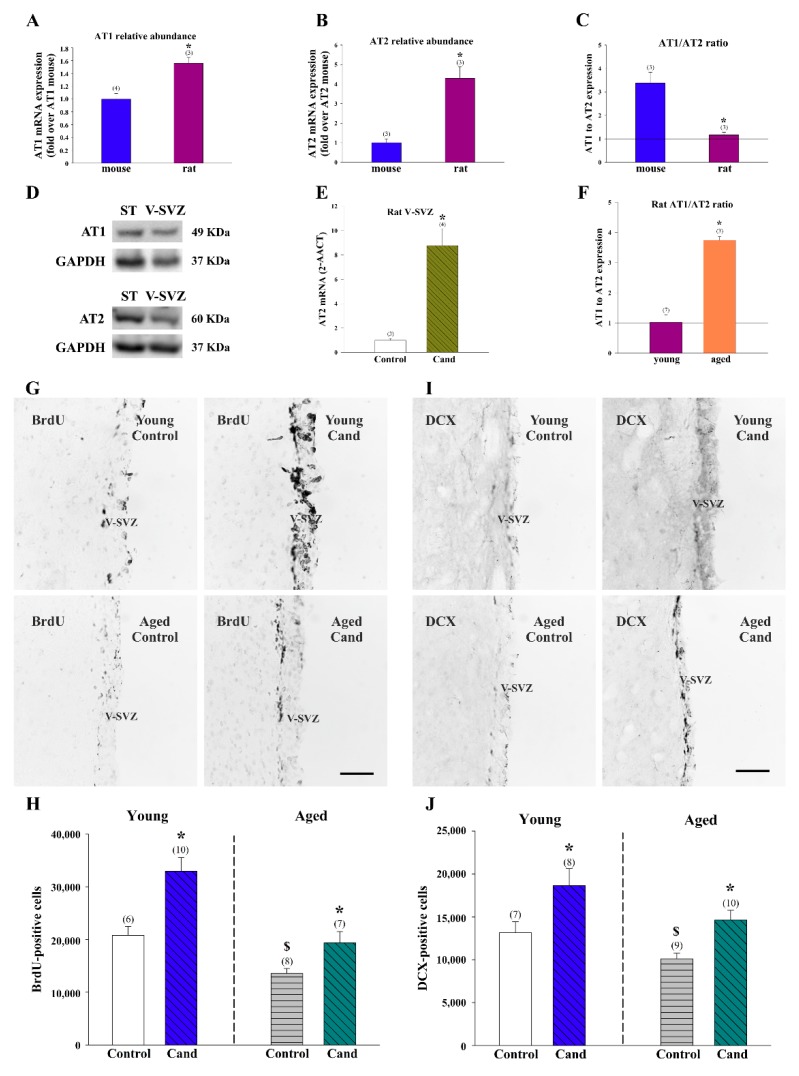
Figure 7.
Expression of AT1 and AT2 receptors in the adult ventricular–subventricular (V-SVZ) of rats and mice. The level of mRNA of AT1 (A) and AT2 (B) was higher in rats than in mice. However, the ratio of AT1/AT2 receptors in the microdissected V-SVZ was higher in mice than in rats (C). (D) Representative bands of AT1 and AT2 receptors in the mouse V-SVZ detected by western blot. Striatum (ST) was used as a positive control. (E) AT2 receptor expression was higher in rats treated with the AT1 receptor antagonist candesartan (Cand) than in control rats. (F) The ratio of AT1/AT2 was higher in aged than in young rats. Representative photomicrographs of coronal sections immunostained for BrdU (G) and DCX (I) of young and aged rat V-SVZ. The estimated number of BrdU and DCX-ir cells in the V-SVZ of the experimental groups is shown in (H,J). Data are means ± SEM. * p < 0.05 relative to the control group of the same age (Student’s t-test); in figures (H,J), $ p < 0.05 relative to the control young group (Student’s t-test). BrdU = bromodeoxyuridine; DCX = doublecortin; SEM = standard error of the mean. Scale bar: 100 μm.

