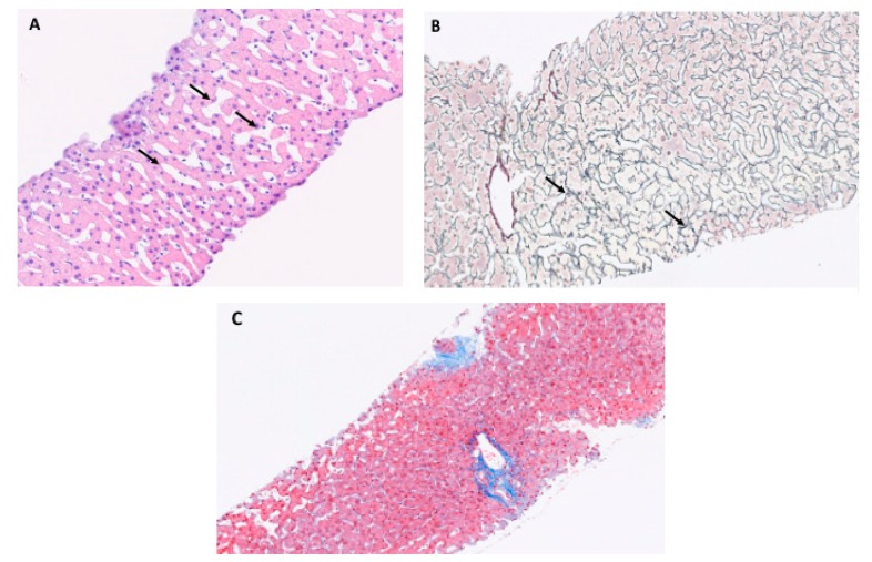
Figure 2.
Histopathology of porto-sinusoidal vascular disease. (A) Section of liver biopsy at 10× magnificiation with hematein eosin with diffuse sinusoidal dilatation (arrows), (B) section of liver biopsy at 10× magnification with argentic reticulin stain with non-fibrous parenchyma preferentially around portal tracts (arrows), (C) Normal liver without signs of porto sinusoidal vascular disease.

