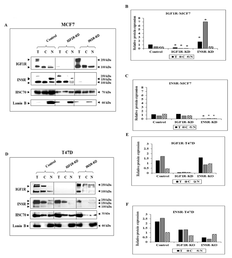
Figure 1.
Subcellular analysis of IGF1R and INSR expression in MCF7-derived IGF1R KD and INSR KD cells and IGF1R/INSR siRNA-transfected T47D cells. (A) MCF7 cells were transfected with shRNAs against IGF1R or INSR (or control shRNA), selected for puromycin resistance, and fractionated into cytoplasmic and nuclear fractions. Western blot analysis of total IGF1R and INSR was conducted using antibodies against total IGF1R and INSR. Lanes with total (T) extracts include 100 μg protein and lanes with cytoplasmic (C) or nuclear (N) extracts include 40 μg protein. Heat shock cognate protein-70 (HSC70) was used as a marker for total protein and lamin B was used as a marker for nuclear fractions. (B,C) Scanning densitometry analysis of basal IGF1R and INSR levels. Bars represent IGF1R and INSR values (AU, arbitrary units), normalized to the corresponding HSC70 levels. Results of an illustrative experiment, repeated twice with similar results, are shown. *, p < 0.01 versus control cells. (D) T47D cells were transfected with siRNA against IGF1R and INSR, or negative control siRNA, and after 72 h IGF1R and INSR were detected by Western blots. (E,F) Optical density is expressed as IGF1R or INSR values normalized to the corresponding HSC70.

