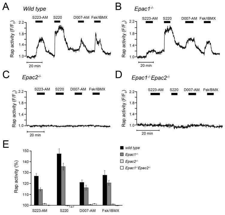
Figure 6.
Changes of plasma membrane Rap activity in primary β-cells from wildtype and Epac-deficient mouse islets. (A) Single-cell TIRF microscopy recording from a wildtype islet transduced with GFP-RalGDSRBD. Representative for 37 cells from five experiments and four independent islet isolations. (B–D) Similar recordings from β-cells isolated from Epac1-/- (B) Epac2-/- (C) and Epac1/2-double knockout mice (D). Representative for 35 (B), 47 (C) and 75 (D) cells from four to five experiments and three independent islet preparations from each genotype. (E) Means ± s.e.m. for the effects of the Epac agonists on Rap activity expressed as time-averaged GFP-RalGDSRBD fluorescence normalized to the baseline.

