Figure 2.
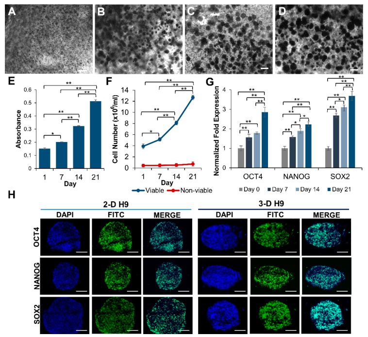
Growth of pluripotent human ESCs in 3-D self-assembling scaffolds. (A–D) Clonal growth of ESCs (H9 cells) encapsulated in PEG-8-SH/PEG-8-Acr scaffolds and incubated in culture medium was observed by light microscopy at 0, 7, 14, and 21 days. (E) Quantitative determination of cell proliferation by 3-(4,5-dimethylthiazol-2-yl)-2,5-diphenyltetrazolium bromide (MTT) assay using microplate reader. Results were expressed as the absorbance ± standard error (SE) with a significant increase in cell number. (F) Growth of ESCs encapsulated in 3-D scaffolds was assayed by direct counts using a hemocytometer, and cell viability was determined by trypan blue exclusion assay at various time intervals. Data presented as cell number (×106 cells/mL) ± SE. (G) Expression of selected pluripotency markers, OCT4, NANOG, and SOX2, in ESCs cultured in self-assembling scaffolds for 0, 7, 14, and 21 days as determined by qRT-PCR. The expression of genes at day 0 was set to 1 and results were expressed as the fold expression ± SE normalized to reference genes HMBS, GAPDH, and β-ACTIN (* p < 0.05 and ** p < 0.01). (H) Confocal images (20X) of 2-D and 3-D grown ESCs displaying the expression of pluripotent proteins, OCT4, NANOG, and SOX2. All scale bars represent 100 μm.

