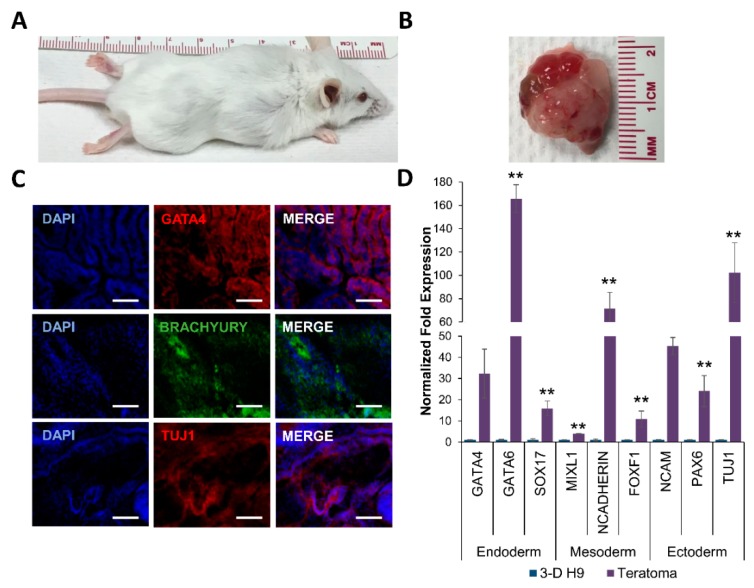
Figure 5.
Teratoma formation by 3-D grown human ESCs in severe combined immunodeficient (SCID) Beige mice. (A) Tumor growth was observed in all mice (n = 3) injected with 3-D grown ESCs (H9 cells). (B) Explanted tumor at 4 weeks showed encapsulated, lobular, and well-circumscribed gross morphology consistent with teratoma growth. (C) Expression of GATA4, BRACHYURY, and TUJ1 proteins representing the endoderm, mesoderm, and ectoderm, respectively in excised teratomas, as shown by confocal images (20X). All scale bars represent 100 μm. (D) Expression of germ layer-specific genes, SOX7 and GATA6 (endoderm), BRACHYURY and MIXL1 (mesoderm), and PAX6 and NCAM (ectoderm) in excised teratomas, as determined by qRT-PCR. Results are expressed as the fold expression ± SE normalized to reference genes HMBS, GAPDH, and β-ACTIN (* p < 0.05 and ** p < 0.01).

