Figure 1.
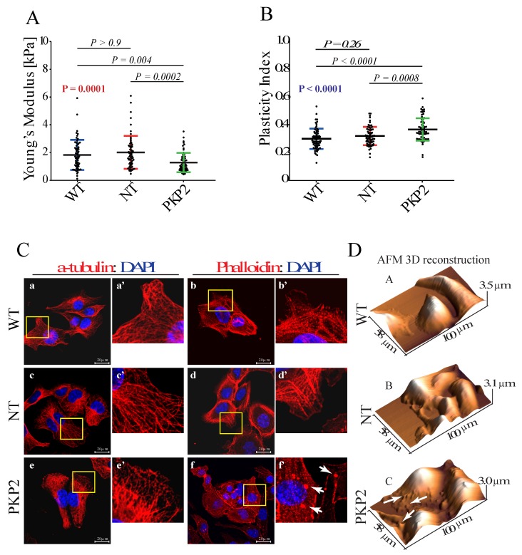
Knock down of PKP-2 affects mechanical properties and morphology of HL-1 cells. (A) Young’s modulus value indicated a reduced stiffness upon knockdown of PKP2 protein. HL-1WT: 1.65 kPa; 0.99 kPa = 25% Percentile; 2.45 kPa = 75% Percentile; HL-1NT: 1.74 kPa; 1.15 kPa = 25% Percentile; 2.48 kPa = 75% Percentile; HL-1PKP2: 1.05 kPa; 0.74 kPa = 25% Percentile; 1.57 kPa = 75% Percentile. n = 60 (179 force curves) for HL-1WT, HL-1NT and (178 force curves) for HL-1PKP2, n = n° of cells. Kruskal–Wallis test with Dunn’s correction, p value in red. (B) Plasticity index showed that in PKP2 knockdown cells external forces are converted more into viscous losses, suggesting a compromised actin network architecture. HL-1WT: 0.30 ± 0.07; HL-1NT: 0.31 ± 0.06; HL-1PKP2: 0.36 ± 0.08. n = 68 (204 force curves) for HL-1WT, n = 67 (201 force curves) for HL-1NT, n = 69 (207 force curves) for HL-1PKP2. One-way ANOVA with Dunnett’s correction, ANOVA p value in blue. (C) Confocal microscopy analysis showed a conserved microtubules structure in all the conditions (panels a, a’, c, c’, e, and e’.), whereas phalloidin staining showed the presence of actin granules in PKP2 knockdown cells (e.g., white arrows in panel f’). n = 3 independent cell staining (scale bar 20 μm). For each IF picture high magnification panels of selected areas are shown. (D) AFM topographic maps confirmed the presence of actin granules, as indicated by arrows in panel c.

