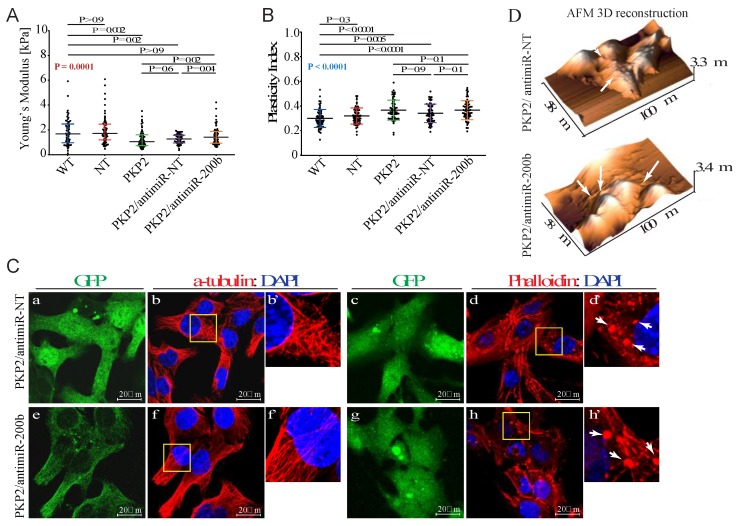
Figure 4.
Downregulation of miR-200b partially rescues mechanical properties of PKP-2 deficient HL-1 cells. (A) Downregulation of miR-200b induces a rescue of the Young’s modulus value in PKP2 knockdown cells. HL-1WT: 1.65 kPa; 0.99 kPa = 25% Percentile; 2.45 kPa = 75% Percentile; HL-1NT: 1.74 kPa; 1.15 kPa = 25% Percentile; 2.48 kPa = 75% Percentile; HL-1PKP2: 1.05 kPa; 0.74 kPa = 25% Percentile; 1.57 kPa = 75%; HL-1PKP2/antimiR-NT: 1.24 kPa; 0.94 kPa = 25% Percentile; 1.56 kPa = 75% Percentile; and HL-1PKP2/antimiR-200b: 1.40 kPa; 0.92 kPa = 25% Percentile; 1.87 kPa = 75% Percentile. n = 60 (179 force curves) for HL-1WT, HL-1NT, and (178 force curves) HL-1PKP2, n = 56 (166 force curves) for HL-1PKP2/antimiR-NT and n = 53 (159 force curves) for HL-1PKP2/antimiR−200, n = n° of cells. Kruskal–Wallis test with Dunn’s correction p value in red. (B) Plasticity index showed that the presence of antagomiR-200b does not rescue the viscoelastic properties in PKP2 knockdown cells. HL-1WT: 0.30 ± 0.07; HL-1NT: 0.31 ± 0.06; HL-1PKP2: 0.36 ± 0.08; HL-1PKP2/antimiR-NT: 0.34 ± 0.07, and HL-1PKP2/antimiR-200b: 0.36 ± 0.07. n = 68 (204 force curves) for HL-1WT, n = 67 (201 force curves) for HL-1NT, and HL-1PKP2/antimiR-NT, n = 69 (207 force curves) for HL-1PKP2, and HL-1PKP2/antimiR-200b. One-way ANOVA with Dunnett’s correction. ANOVA p value in blue. (C) Confocal microscopy analysis showed a conserved microtubules structure after antimiR expression (panels b, b’, f, and f’). Phalloidin staining showed the partial recovery of F-actin by antimiR-200b expression, whereas actin granules were still present (white arrows in panels d, d’, h, and h’). GFP channel (panels a, c, e, and g) was used as antagomiRs transduction control. n = 3 independent cell staining (scale bar 20 μm). (D) AFM topographic images confirmed the presence of actin granules also after antimiR-200b expression (arrows in panels).

