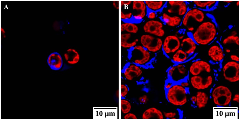
Figure 4.
Confocal microscopy images of Chlamydomonas reinhardtii cc124 under normal condition (A) and under salt stressed (150 mM NaCl) condition (B). Calcofluor white stains cellulose and chitin and is blue in color while photosystem II was excited and visualized in red. These images show an increase in polysaccharides as an integral event of the palmelloid formation (unpublished in-house data).

