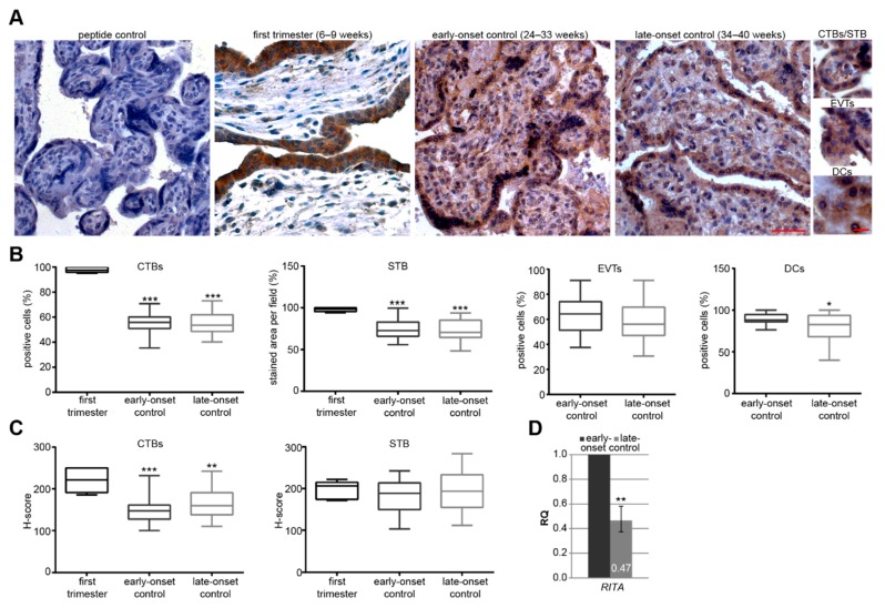
Figure 1.
RBP-J (recombination signal binding protein J)-interacting and tubulin-associated protein (RITA) is expressed in trophoblastic cells of placental tissue and its gene expression decreases at late stages of gestation. (A) Paraffin-embedded tissue sections were immunohistochemically stained with a specific RITA antibody (brown) and counterstained with hematoxylin (blue). RITA antibody neutralized with its corresponding peptide (peptide control) was used as negative control. Scale of images: 50 µm, scale of small images: 20 µm. CTBs, cytotrophoblasts; STB, syncytiotrophoblast; EVTs, extravillous trophoblasts; DCs, decidual cells. (B) Quantification of RITA positive cells in first trimester placental sections (6–9 weeks, n = 6), early-onset control (24–33 weeks; n = 20), and late-onset control samples (34–40 weeks; n = 21). The results are presented as box and whisker plots with minimum and maximum variations. Student’s t-test, * p < 0.05, ** p < 0.01, *** p < 0.001. (C) Semi-quantitative analysis of the RITA staining using the H-score method. The results are presented as box and whisker plots with minimum and maximum variations. Student’s t-test referring to first trimester samples, ** p < 0.01, *** p < 0.001. (D) The relative amount of the gene RITA was analyzed from placental tissues from late-onset (n = 17, 34–40 weeks) compared to early-onset controls (n = 13, 26–33 weeks). The results are presented as relative quantification (RQ) with minimum and maximum range and statistically compared between both groups. Student’s t-test, ** p < 0.01. The mean value of the expression levels of succinate dehydrogenase complex, subunit A (SDHA), TATA box-binding protein (TBP), and tyrosine 3-monooxygenase/tryptophan 5-monooxygenase activation protein, zeta polypeptide (YWHAZ) was used as endogenous control. Clinical information is listed in Table 1.

