Figure 8.
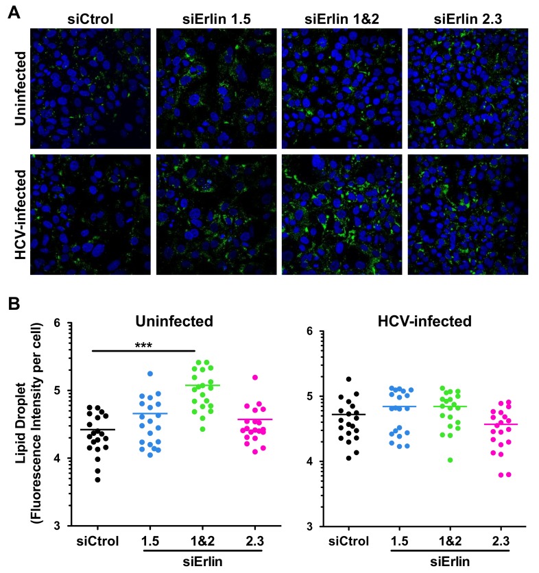
Erlin-1 protein down-regulation increases LD content in Huh-7 cells. siRNA-transfected Huh-7 cells were inoculated with JFH-1 D183 virus at high multiplicity of infection (moi = 3) and the intracellular LD content was analyzed 48 h after infection as described in Material and Methods. (A) Representative confocal image sections of intracellular LD accumulation (in green) in siRNA-transfected HCV-infected and uninfected cells. Nuclei (in blue) were counterstained with Hoechst dye. (B) Quantitation of the total LD fluorescence intensity signal per cell from confocal image sections. ImageJ software was used to quantitate the total LD fluorescence intensity signal in twenty individual cells of each condition. Each dot in the graph represents the LD fluorescence intensity of an individual cell and the horizontal lines show the average LD fluorescence intensity in each group of data. One-way ANOVA followed by Dunnett’s Multiple Comparison Test was used to determine the statistical significance (*** p < 0.001).

