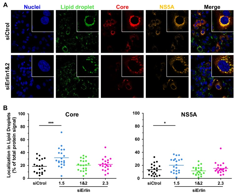
Figure 9.
Erlin-1 protein down-regulation does not impair the localization of HCV core and NS5A proteins surrounding LDs. siRNA-transfected Huh-7 cells were inoculated with JFH-1 D183 virus at high multiplicity of infection (moi = 3) and the intracellular HCV protein and LD content was analyzed as described in Material and Methods. (A) Representative confocal images of the intracellular localization of HCV core (in red) and NS5A (in orange) proteins surrounding LDs (in green) in ERLIN down-regulated (siErlin 1&2) and control (siCtrol) HCV-infected cells. Nuclei (in blue) were counterstained with Hoechst dye. The panels on the right side show merged images of the four channels of each image. White boxes in the upper right side of each panel show zoom-in images of single cells for a more detailed observation. (B) The fluorescence intensity signal of core and NS5A proteins associated to LDs was quantitated in twenty randomly selected HCV-infected cells of each condition. Each dot in the graph represents the percentage of the fluorescence intensity signal of core or NS5A protein associated to LDs in a given cell, while the horizontal lines show the average of all data points in each group. One-way ANOVA followed by Dunnett’s Multiple Comparison Test was used to determine the statistical significance (* p < 0.05; *** p < 0.001).

