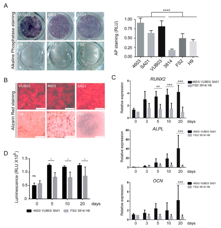
Figure 1.
Heterogeneity in osteogenic differentiation potential of mesodermal progenitor cells (MPCs) derived from human pluripotent stem cell (hPSC-MPC) lines. (A) Comparison and quantification of alkaline phosphatase staining in MPCs derived from six hPSC lines, VUB03, 4603, SA01, H9, 3814, and FS2, cultured for ten days in osteogenic differentiation medium. Data obtained from 3 independent experiments are represented as the mean ± SD. Statistical analysis was based on an unpaired t-Test; **** p < 0.0001. (B) Comparison of alizarin red staining in MPCs derived from six hPSC lines, cultured for twenty days in osteogenic differentiation medium. Data correspond to 3 independent experiments (Scale bar: 500 µM). (C) Time-course analysis of RUNX2, ALPL, and OCN gene expression by quantitative RT-QPCR analysis in VUB03, 4603, and SA01 MPCs versus H9, 3814, and FS2 MPCs. Data obtained from independent experiments for the 6 hPSC lines are represented as the mean ± SD. (D) Time-course analysis of viable cell number evaluated by the ATP content during osteogenic differentiation of MPCs VUB03, 4603, SA01 compared to MPCs H9, 3814, FS2 with poor differentiation potential. Data obtained from 3 independent quantifications for each cell line are represented as the mean ± SD. For C and D, statistical analysis was based on a two-way ANOVA followed by a Sidak’s multiple comparison. * p < 0.05, ** p < 0.01, *** p < 0.001, ns: not significant.

