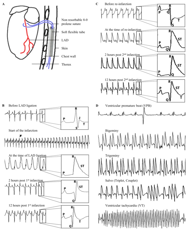
Figure 1.
Induction of ventricular arrhythmias (VA). Schematic drawing of the loose left anterior descending (LAD) ligature positioning (A). Mouse ECG changes in relation to the time of the firs LAD ligation during ischemia-reperfusion infarction (B). Mouse ECG changes in relation to the time of the permanent second LAD-ligation (reinfarction and URI group C). Mouse ECG strips showing different types of observed VA (D).

