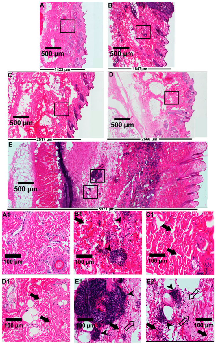
Figure 7.
Microscopic evaluation of rabbit skin exposed to recombinant toxins (LiRecTCTP, LiRecDT1, or combined toxins LiRecTCTP/LiRecDT1). Light microscopic analysis of tissue sections was performed on rabbit skin after 24 h of injection. The tissue sections were stained with hematoxylin and eosin. Edema triggered in rabbit skin by (A) GFP, (B) LiRecDT1 (1 µg), (C) LiRecTCTP (10 µg), (D) LiRecTCTP (20 µg), and (E) the combination of LiRecDT1 (1 µg) and LiRecTCTP (20 µg), as visualized by skin thickness. Skin structures are compared via scanning of images from epidermal (on the right of figure) to muscular tissues (on the left of figure) under the same laboratory conditions (Scale bars indicate 500 µm). The width of the tissue (E) points to a deep edema after LiRecTCTP and LiRecDT1 combination when compared to toxins alone (B, D). Isolated LiRecTCTP (C, D) induced a higher edema compared to LiRecDT1 alone (B) or negative control GFP (A), which shows a normal skin histology (A1). An intense inflammatory response with the presence of neutrophils and fibrinoid exudates into the dermis is shown when both toxins were administered (E1, E2) compared to isolated LiRecTCTP (C1, D1) or LiRecDT1 (B1) (Scale bars indicate 100 μm). Closed arrows indicate disorganization of collagen fibers and dermal edema, closed arrowheads indicate a massive inflammatory response with the presence of neutrophils, and open arrows indicate fibrin network deposition. Thickness of the skin tissue (A–E) is shown in the bottom of each tissue section (µm).

