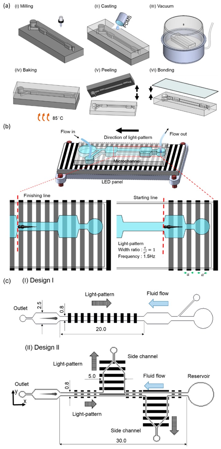
Figure 1.
Fabrication and experimental system. (a (I–VI)) Fabrication of the microfluidic device consisting of two microwells and one experimental chamber. (b) Experimental setup consisting of a microfluidic, flow control, and optical illumination setup platform. The optical setup illuminated the larvae from the bottom with an optimum grating frequency of 1.0 Hz and a grating width ratio () of 1:1. A syringe pump was connected at the outlet to draw fluid, while the fluid was introduced manually at the inlet. The schematics are not to scale. (c) Design and dimensional details of both of the microfluidic devices used for the experiments. All dimensions are in mm.

