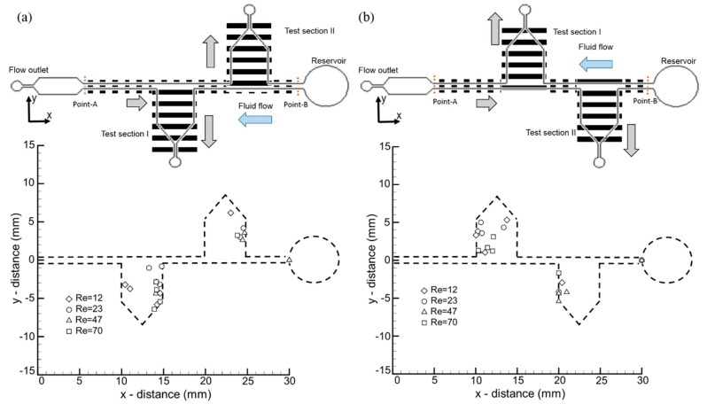
Figure 5.
The modified microfluidic channel with two additional test sections, namely test section I and test section II, where the optical stimulus was provided according to the channel opening. The location of the final resident point of the individual zebrafish larvae is depicted in 2D coordinates under different flow conditions. The symmetric and direction-specific nature of larvae migration was tested through a microfluidic device with test section I on the (a) right-hand and (b) left-hand side of the larvae’s swimming direction.

