Figure 1.
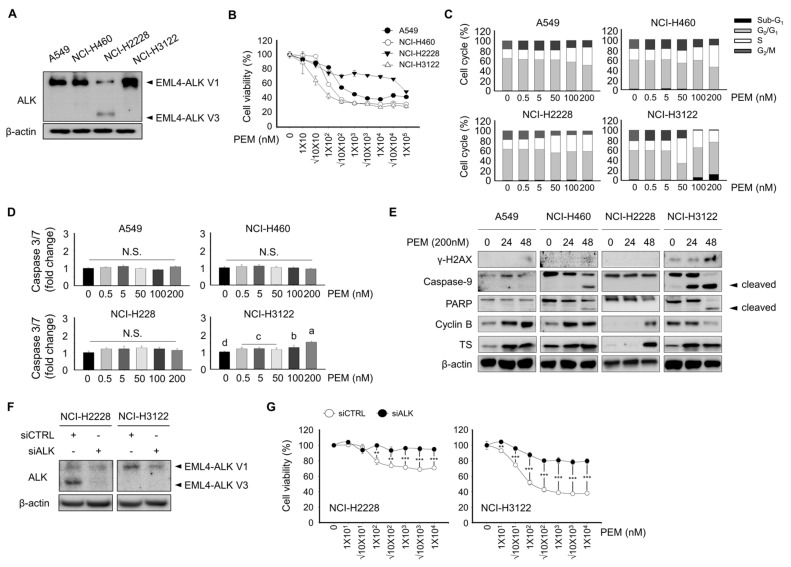
Effect of pemetrexed on cell viability, cell cycle, and apoptosis in lung cancer cell lines. (A) A549, NCI-H460, NCI-H2228 EML4-ALK V3, and NCI-H3122 EML4-ALK V1 cells were evaluated for their basal expression of ALK protein by Western blotting; β-actin served as the loading control. (B) Cell viability assay. A549, NCI-H460, NCI-H2228, and NCI-H3122 cells were treated with the indicated concentrations of PEM for 3 days. (C) Cell cycle analysis by PI staining and flow cytometry. A total of 1 × 10⁶ cells were seeded in 60-mm plates and treated with 0, 0.5, 5, 10, 50, 100, and 200 nM of PEM for 24 h. Data are presented as histograms (black, Sub-G1; gray, G0/G1 phase; white, S phase, and dark gray, G2/M phase). (D) Caspase 3/7 activity was quantified 24 h after PEM treatment in A549, NCI-H460, NCI-H2228, and NCI-H3122 cells. Means with different letters (a, b, c, and d) indicate statistically significant differences (p < 0.05). (E) γ–H2AX, Caspase-9, PARP, cyclin B, and TS expression in A549, NCI-H460, NCI-H2228, and NCI-H3122 cells, as determined by Western blotting; β-actin served as the loading control. (F) Expression of ALK was evaluated using Western blotting in NCI-H2228 and NCI-H3122 with or without siALK; β-Actin served as the loading control. (G) Cell viability assay. NCI-H2228 and NCI-H3122 cells with or without siALK were treated with the indicated concentrations of PEM for 3 days.

