Figure 2.
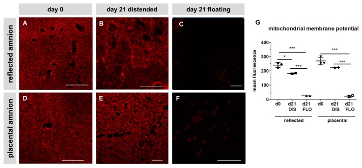
Mitochondrial membrane potential of reflected (A,B,C) and placental (D,E,F) amnion. Mitochondrial membrane potential (red) was stained with tetramethylrhodamin-methylester (TMRM; 500 nM) at day 0 (A,D) and at day 21 in biopsies cultivated while mechanically stretched (DIS; B,E) or kept floating (FLO; C,F). Imaging was performed with an inverted confocal microscope (LSM510, Zeiss, excitation/emission 543 nm/585 nm). Image analysis was performed with Zeiss ZEN2009 software (G). Mean ± standard deviation (SD), n = 3 (donors); representative images of one donor. Scale bar: 100 µm. DIS: distended biopsies; FLO: floating biopsies. Level of significance is indicated as *p < 0.05, ***p < 0.001

