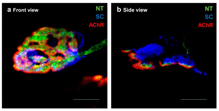
Figure 1.
Cellular components of the neuromuscular junction (NMJ). Representative confocal micrographs of healthy NMJs from levator auris longus muscle showing a NMJ in a front view in (a) and an NMJ in side view in (b). The synapses are multiply immunofluorescent-stained: SNAP-25 in green to stain the nerve terminal (NT); S100 in blue to stain the Schwann cells (SC) and AChRs in red to stain the postsynaptic membrane. Scale bars = 10 μm.

