Figure 3.
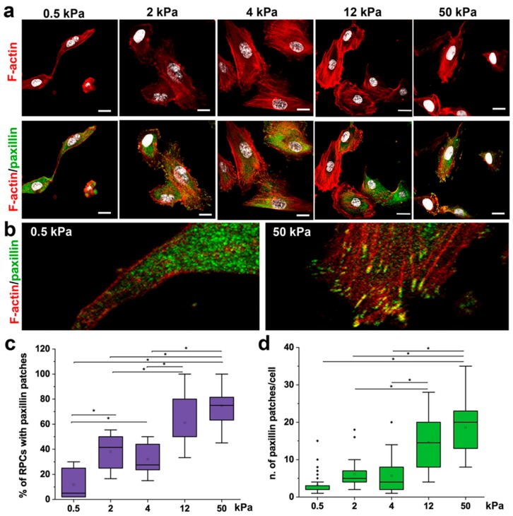
Substrate stiffness modulates cytoskeleton organization and FA formation. (a) Confocal images of F-actin immunodetection by phalloidin (red), paxillin (green) and nuclei with DAPI counterstain (white) of RPCs cultured on substrates with different stiffness. F-actin organization shows a trend, from diffuse on soft gels to progressively organized on stiffer substrates (as stress fibers). (b) Higher magnification images showing that paxillin staining was diffuse on soft substrate (left), or organized in clusters on the cell membrane in stiff conditions (right). (c) Percentage of RPCs containing paxillin clusters in function of stiffness. At least 10 representative images from each condition were analyzed. (d) Average number of paxillin patches in cell cultured on different stiffness. At least 20 cells for each condition were analyzed. Box-and-whisker plots: line = median, box = 25–75%, whiskers = 10–90%. *p < 0.05 using one-way ANOVA followed by Tukey’s post-hoc test. Bars = 25 µm.

