Figure 1.
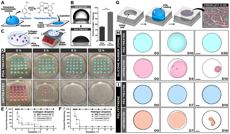
(A) Surface modification of PDMS is achieved simply by incubating the surface with a dopamine hydrochloride solution to form a thin surface coating. (B) PDA-functionalized PDMS surface shows decreased water contact angle as compared to untreated PDMS. (C) The effect of PDA coating was tested by shaking hydrogel droplets formed on PDA-treated or untreated PDMS surfaces that were immersed in phosphate-buffered saline to prevent dehydration. (D) The hydrogel droplets on the PDA-treated PDMS remain stable for 12 h of constant agitation, whereas the droplets rapidly detach from the untreated PDMS surface. The quantification of remaining (E) collagen and (F) matrigel droplets at different time points shows significantly improved hydrogel adhesion due to PDA surface coating. (G, H) Human lung fibroblasts (hLFs) cultured in type I collagen hydrogel remain attached to PDA-treated PDMS for prolonged periods but begin to detach from untreated PDMS wells after 4 days. The hydrogel constructs were pseudo-colored in the presented images. The dotted lines indicate the outlines of culture wells. Scale bars: 2 mm. (I) Similar beneficial effects of PDA are observed in the 3D culture of hLFs in polystyrene wells. Scale bars: 2 mm. ***P < 0.001. Data are expressed as mean ± SEM (n = 6).

