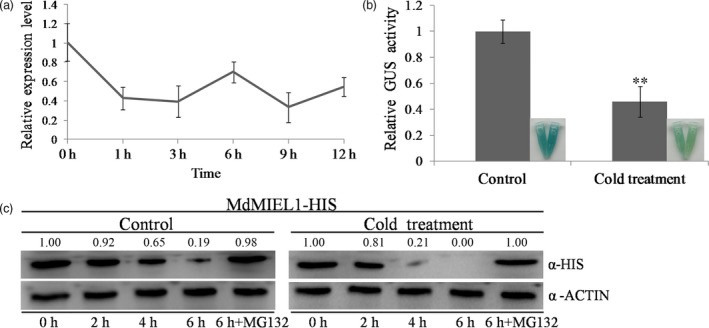
Figure 8.

The expression pattern of MdMIEL1 in response to cold stress (4°C treatment). (a) MdMIEL1 gene expression detected by qRT‐PCR analysis. Experiments were performed in three biological replicates and three technical replicates. The value for 0 h was set to 1. (b) GUS staining and relative GUS activity analysis of the MdMIEL1 promoter expression construct ProMdMIEL1::GUS in transgenic apple calli. Control: GUS staining and activity analysis at 9 h at 24°C. Cold treatment: GUS staining and activity analysis at 9 h under cold stress. (c) Degradation of the MdMIEL1‐HIS fusion protein under cold stress. Total proteins extracted from wild‐type apple calli with or without 4°C treatments and the inclusion of 100 μm MG132 were incubated with the purified MdMIEL1‐HIS fusion protein. The samples were collected at the indicated time. Control: 24°C; cold treatment: 4°C. ACTIN was used as internal reference. The relative intensity ratio between the HIS and the ACTIN was shown. Each experiment was performed in three replicates. Error bars denoted standard deviation. Significant differences were detected by t‐test (**P < 0.01).
