Figure 2.

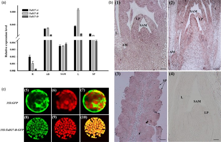
Expression patterns of TaD27s and subcellular localization of the TaD27‐B protein. (a) Expression patterns of TaD27s in wheat. R, roots (Z12); AB, axillary buds (Z15); SAM, shoot apical meristem (Z13); L, leaves (Z13); SP, spikelet primordia (Z17). (b) In situ hybridization analysis of TaD27‐B. (1) Longitudinal section of the shoot apex at the single ridge stage showing axillary buds (Z13). (2) Longitudinal section of the shoot apex at double ridge stage showing axillary buds (Z15). (3) Longitudinal section of a spike (Z17). (4) Sense probe control; AM, axillary meristem, SP: spike primordium. Bars = 100 μm. (c) Subcellular localization of the TaD27‐B protein. (5–7) The pROKII control vector is located in the cell membrane and cytoplasm; (8–10) 35S::TaD27‐B‐GFP is located in the chloroplasts. Bars = 5 μm.
