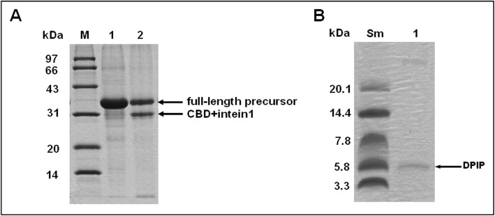
Fig. 5.
DPIP is released from CI-DPIP after intein cleavage. a: Cleavage of the DPIP fusion protein under the optimized condition. M: MW marker (97, 66, 43, 31, 24, 14 kDa). Lane 1: the chitin resin after protein binding. Lane 2: the resin after intein cleavage. The fractions were analyzed by 12% SDS-PAGE. b: DPIP eluted from resin. M: MW marker (20.1, 14.4, 7.8, 5.8, 3.3 kDa). Lane 1: DPIP eluate. The fractions were analyzed by 16.5% Tricine-SDS-PAGE

