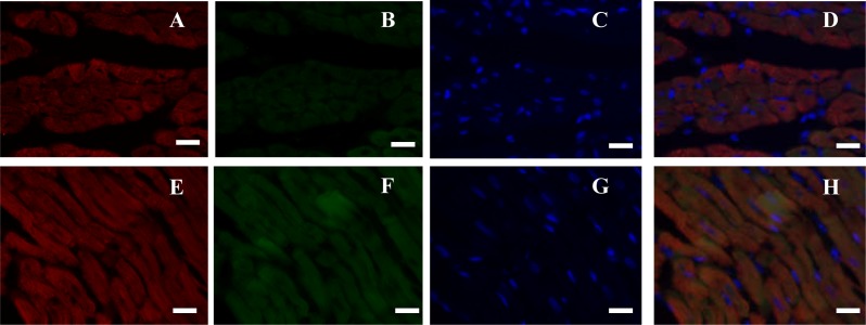
Figure 5. Double immunofluorescence (IF) analysis of rat heart (bar = 20 µm).
(A–D) Cardiac muscle fibers in transection; (E–H) cardiac muscle fibers in longitudinal section. Red fluorescence was stained to locate Myl3 by Alexa Fluor® 647-linked secondary antibody, green fluorescence was stained to locate NeuN by FITC-linked secondary antibody, and blue fluorescence was stained to locate nucleus by Hoechst 33342.

