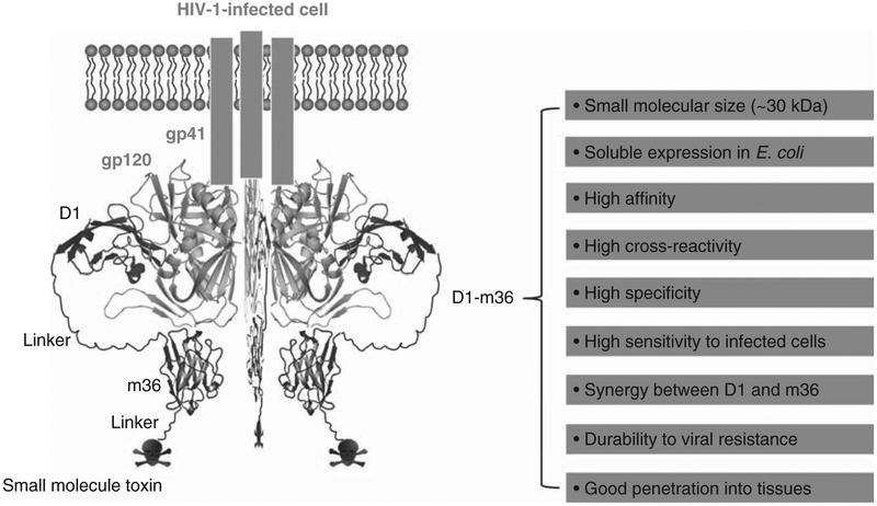
Figure 3. Schematic representation of specific targeting of potentially all HIV-1-infected cells by D1–m36–small molecule toxin conjugates.
The X-ray crystal structures of HIV-1 gp120 and D1 were adapted from PDB entry 1GC1. The m36 structure is derived from homology modeling using SWISS-MODEL (http://swissmodel.expasy.org/) based on the crystal structure of a human VH domain (PDB entry 1T2J). m36 was manually docked into the CoRbs of gp120 according to the crystal structure of D1D2–gp120–17b complex (PDB entry 1GC1). Favorable properties that the D1–m36 combination could exhibit are shown on the right.

