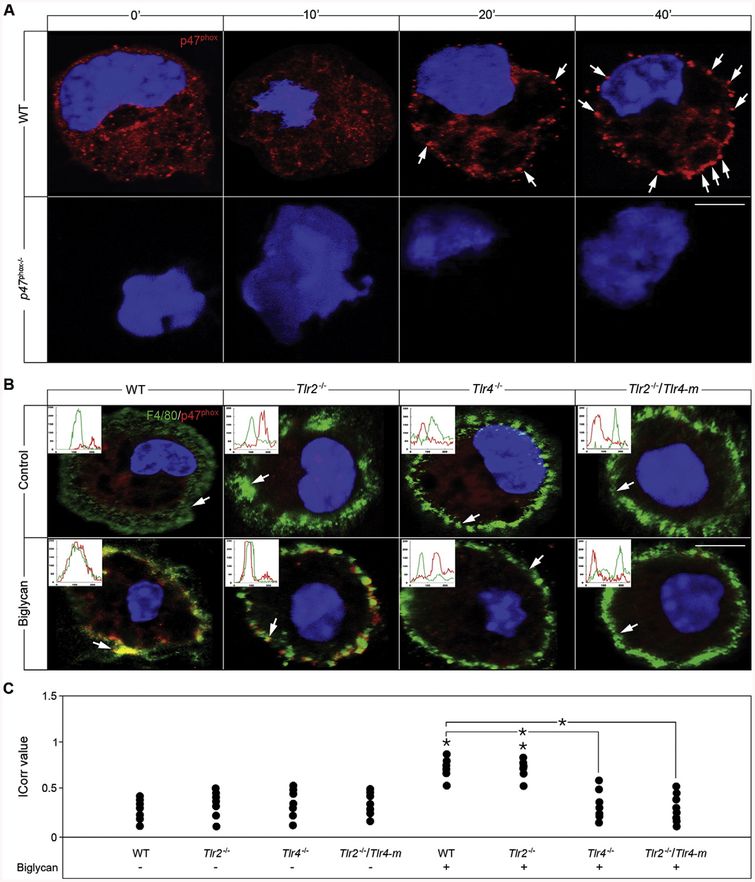
Fig. 4.
Biglycan triggers p47phox translocation in a TLR4-dependent manner. (A) Confocal images of p47phox (red) in macrophages isolated from WT mice either untreated or stimulated with biglycan (4 μg/ml) for 10, 20 and 40 min. White arrows indicate localization of p47phox (red) to the plasma membrane. Nuclei were stained with DAPI (blue). (B) Confocal images of F4/80 (green) and p47phox (red) in macrophages isolated from WT, Tlr2−/−, Tlr4−/− and Tlr2−/−/Tlr4-m mice either untreated or after 40 min treatment with biglycan (4 μg/ml). Inserts in the confocal images represent line-scanned profiles, while the white arrows indicate the distance within which the scans were generated. Bar = 10 μm; *P < 0.05. (C) Quantification of p47phox co-localization with F4/80 from (B) given as dot plot for the mean correlation index (Icorr) ± S.D. of at least 10 cells per condition.

